Academic Conference
Application | CNN4 and IncV3 Deep Learning Algorithms in Automated p16/Ki-67 Dual-Stain Cytology Diagnosis and Heer Medical AI Dual-Stain Application Solution
Introduction to the application of CNN4 and IncV3 deep learning algorithms in automated p16/Ki-67 dual-stain cytology diagnosis
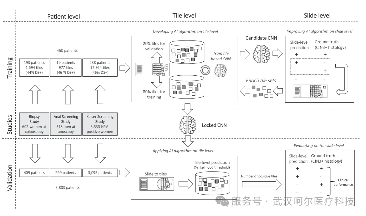
First, the authors selected digital slides from 450 patients from the CYTOREADER cloud platform (Google) as a training set. The slides were cut into tiles of the same size (384x384 pixels, 20X), with 80% of the tile samples selected for initial algorithm training and 20% for algorithm validation.
The CNN4 and IncV3 algorithms were used to detect the number of tiles exceeding a specific likelihood threshold, thereby determining the number of dual-stain positive cells on the slide. The algorithm provides a probability threshold for each tile (0.5 for CNN4, 0.4 for IncV3), and tiles exceeding this threshold are considered positive results.
The candidate CNN models obtained from initial training were applied at the whole-slide level, and the positive status of slides was determined based on the number of positive tiles (CNN4 >= 3 tiles, IncV3 >= 2 tiles). For misclassified slides, false positive or false negative patches were extracted and fed back into the original CNN training to optimize classification accuracy. The final locked CNN models were applied to a blinded validation set containing 3,803 slides.
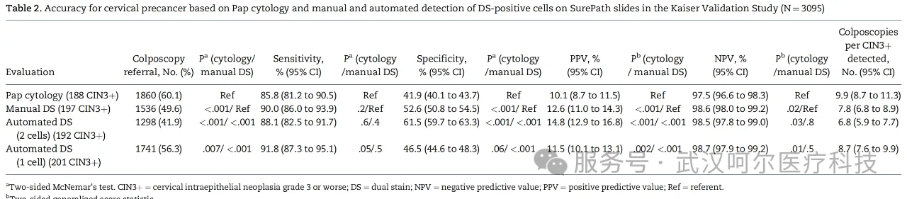
The results showed that in the cervical biopsy validation set (409 cases), CNN4 achieved an AUC of 0.74, sensitivity of 87.0%, and specificity of 45.6%. Among 3,095 HPV-positive women, AI-assisted dual-stain (cutoff >= 2 positive cells) achieved a sensitivity of 88.1% and specificity of 61.5%, significantly outperforming Pap smear (sensitivity 85.8%, specificity 41.9%) and manual dual-stain (sensitivity 90.0%, specificity 52.6%). AI-assisted dual-stain reduced the colposcopy referral rate from 60.1% for Pap smear to 41.9% (P<0.001), and the number of colposcopies needed per CIN3+ detected decreased from 9.9 to 6.8.
Automated digital scanning evaluation eliminates the residual subjective judgment in cervical cancer screening, bringing consistent and stable testing quality to healthcare providers and patients. The transition from traditional Pap smear testing to automated digital scanning technology not only significantly reduces the number of colposcopies but also demonstrates outstanding detection performance in simulated fully vaccinated populations. Through cloud deployment, this technology has global accessibility. The study shows that AI technology not only achieves automation and objectivity but also brings substantial benefits to women's health by reducing unnecessary colposcopy examinations.
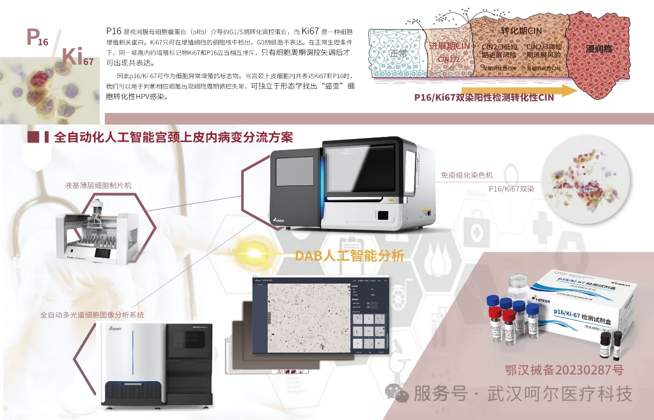
Heer Medical's self-developed p16/Ki-67 dual-stain detection supports the full-process triage solution for cervical cancer screening. The combination of p16/Ki-67 dual-stain and DNA ploidy quantitative detection yields CIN2+ sensitivity, specificity, and diagnostic concordance rates of 92.9%, 71.4%, and 86.3%, respectively, with the diagnostic concordance rate significantly higher than that of DNA ploidy analysis alone [2]. The combined detection can effectively improve accuracy and can be used as an auxiliary tool for ASCUS triage diagnosis.
References
[1]Wentzensen N, et.al. Accuracy and Efficiency of Deep-Learning-Based Automation of Dual Stain Cytology in Cervical Cancer Screening. J Natl Cancer Inst. 2021 Jan 4;113(1):72-79.
[2]Yin L, Ma CB, Liu P, et al. Application value of P16/Ki-67 dual-stain combined with DNA ploidy analysis in ASCUS triage diagnosis[J]. Carcinogenesis, Teratogenesis and Mutagenesis, 2020, 32(04):264-268.
Wuhan Heer Medical Technology Development Co., Ltd.

Wuhan Heer Medical Technology Development Co., Ltd., established in 2006, is a wholly-owned subsidiary of Boai Xinkaiyuan Medical Technology Group Co., Ltd. (stock code: 300109). Heer Medical reshapes the pathology ecosystem with "digital foundation + intelligent hub." The company's AI tumor cell detection technology platform integrates imaging, analysis, and auxiliary reagents into one, providing an integrated solution that significantly improves the efficiency and accuracy of pathology diagnosis.