Academic Conference
Literature Review | AI-Assisted Cervical Cancer Screening Model
Literature review: Research on the application of artificial intelligence models in assisted cervical cancer screening
Cervical cancer is a common gynecological malignancy that threatens women's health, and it is expected to be eliminated through a mature and effective three-level prevention strategy. However, current cervical cancer screening technologies still face bottlenecks, and there is an urgent need to explore new, accurate, efficient, and economically adaptable screening methods. With the continuous development of artificial intelligence (AI) technology, numerous studies have explored the feasibility and practicality of applying deep learning-based AI technology in assisted cervical cancer screening.
This article presents the AI-assisted diagnostic system for rapid TBS classification of cervical liquid-based cytology smears (AIATBS system), described in the paper "Hybrid AI-assistive diagnostic model permits rapid TBS classification of cervical liquid-based thin-layer cell smears." This paper was jointly published in Nature Communications by multiple institutions including the Department of Pathology at Nanfang Hospital of Southern Medical University, Shenzhen Bao'an People's Hospital, the Agency for Science, Technology and Research (A*STAR) Singapore, and the Bioinformatics Institute.
Original article link: https://www.nature.com/articles/s41467-021-23913-3
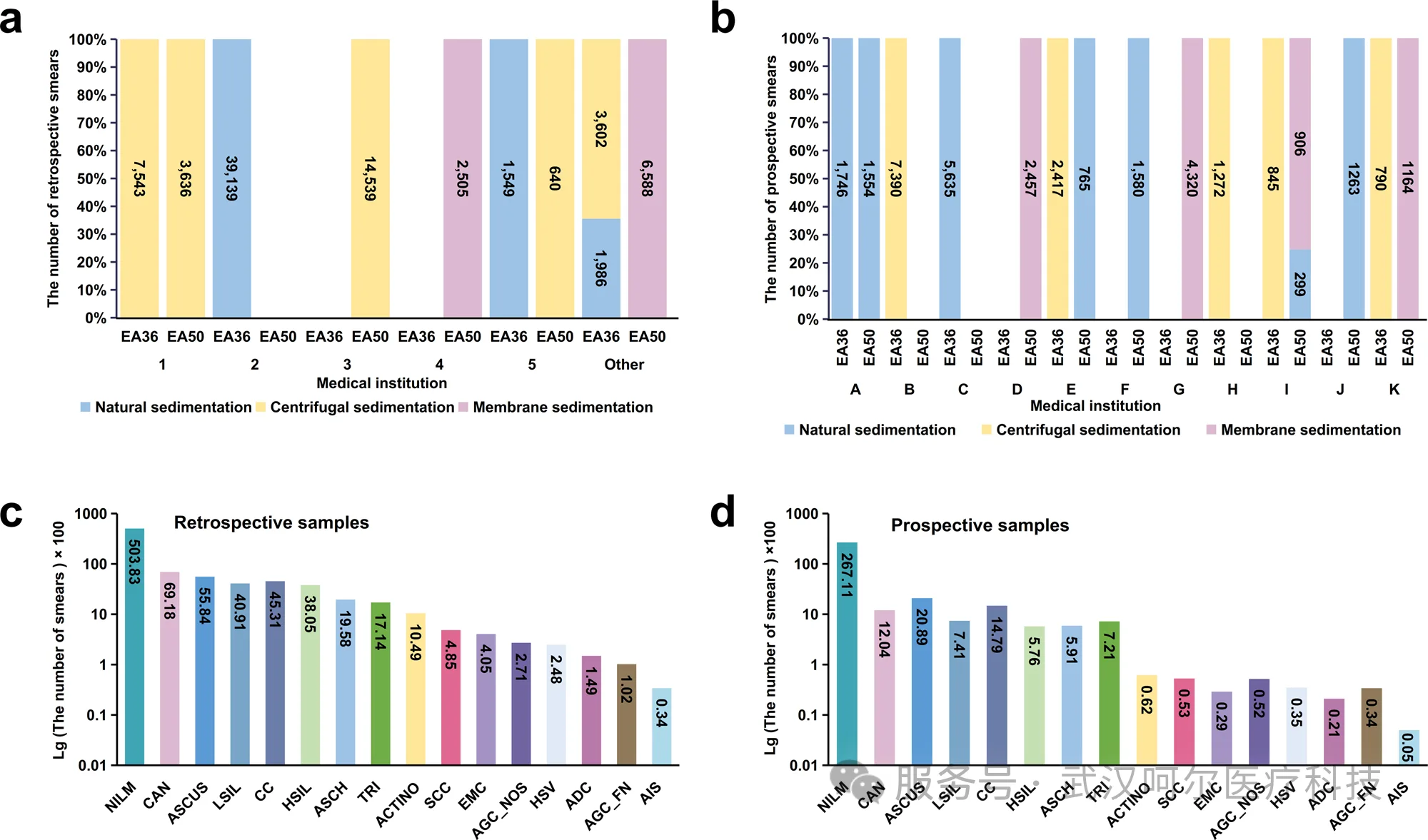
Retrospective data came from 5 medical institutions, including natural sedimentation, centrifugation, and membrane-based preparation methods, as well as two staining methods (EA36 and EA50), totaling 81,727 cell smears. Prospective data came from 11 medical centers with the same types, comprising 34,403 cell smears.
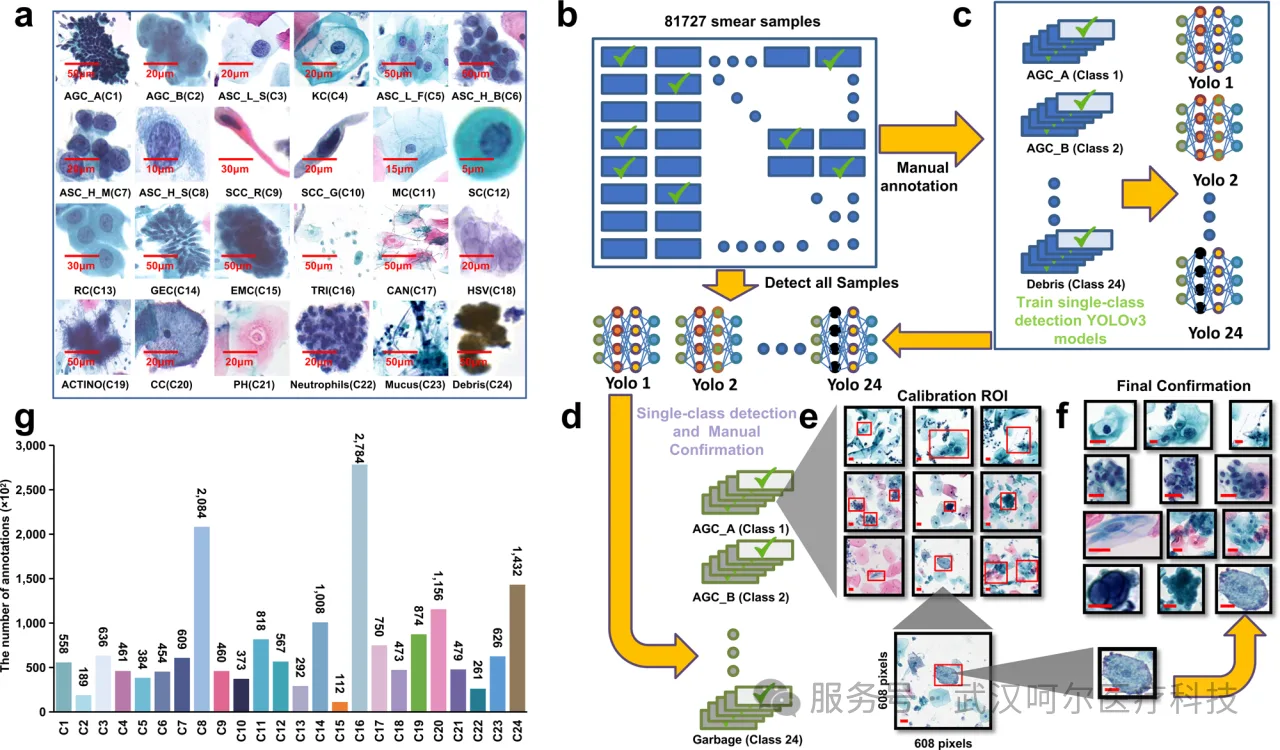
- First, annotation types were categorized into 24 classes based on TBS classification: AGC-A, AGC-B, ASC-L-S, KC, ASC-L-F, ASC-H-B, ASC-H-M, ASC-H-S, SCC-R, SCC-G, MC, SC, RC, GEC, EMC, TRI, CAN, HSV, ACTION, CC, PH, Neutrophils, Mucus, Debris.
- YOLOv3 was used to train 24 single-class models (each model trained with 2,000 annotated data points).
- Manual review of the annotation and recognition results from the 24 classification models on 81,727 samples.
- Finally, three pathology experts confirmed the accuracy of the final annotated data, thus completing the semi-supervised annotation model training.
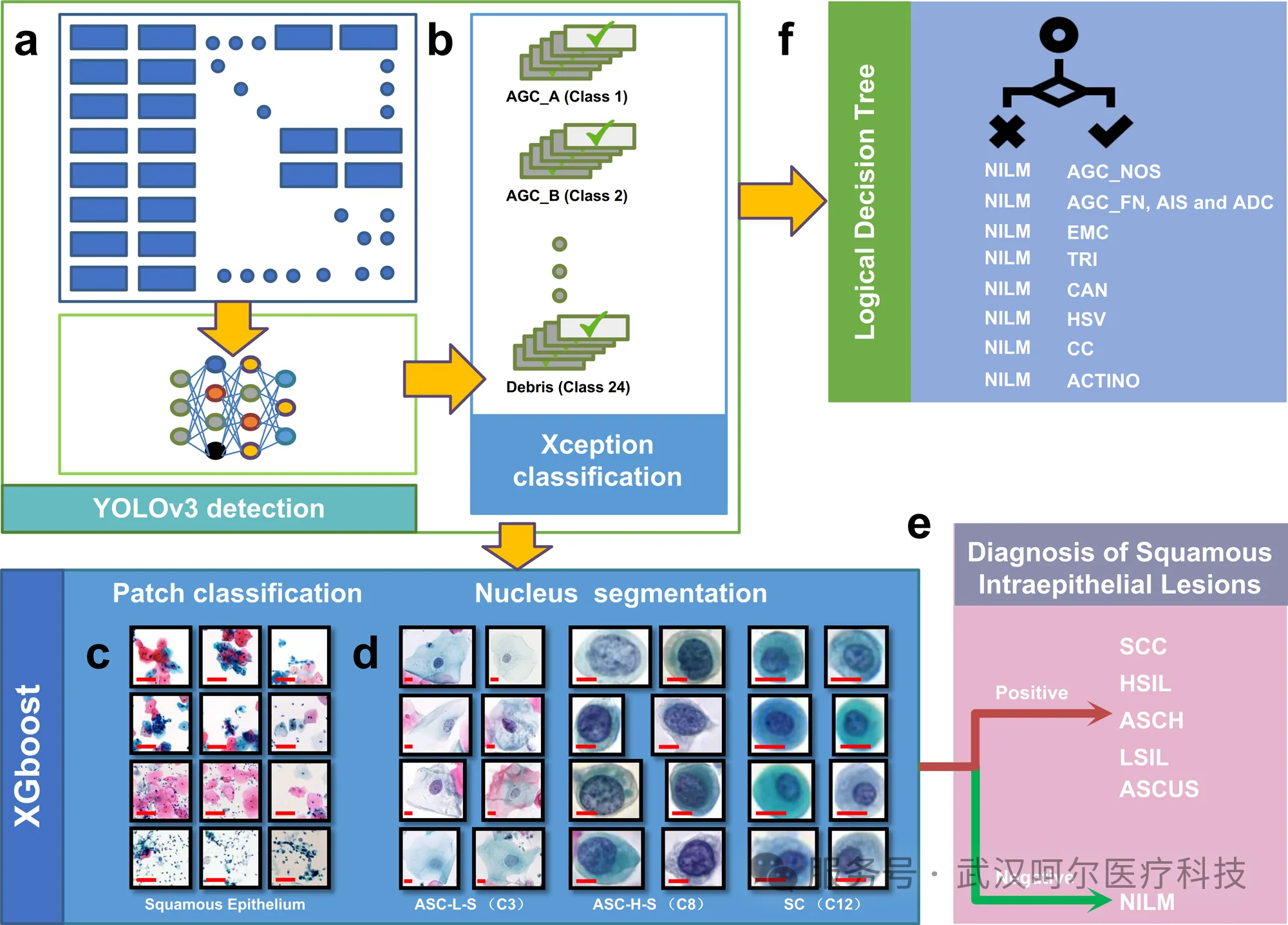
- The YOLOv3 model detects all retrospective samples and extracts detection targets and probabilities.
- The Xception model predicts targets detected by the YOLOv3 model, then extracts classification information and probability data.
- The Patch model extracts regional classification and squamous epithelial target probabilities.
- The UNet nuclear segmentation model classifies cells based on nuclear parameters.
- The XGBoost model is used to predict squamous epithelial lesions.
- The logical decision model is used for classification of glandular epithelial lesions, endometrial cells, and microbial infections.
04AIATBS Model Technical Pipeline
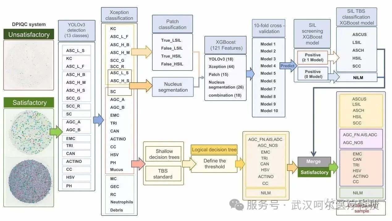
AIATBS integrates multiple deep learning models. First, the DPIQC quality control system performs sample satisfaction detection. Then the YOLOv3 object detection model detects pathological targets, merging annotation categories with relatively consistent pathological types to improve YOLOv3 learning capability and detection efficiency. The detected targets from YOLO are input into the Xception model for feature extraction and classification. The Patch classification model and UNet nuclear segmentation model are further combined to build XGBoost model parameters for detecting squamous epithelial lesions. The XGBoost model (for detecting squamous intraepithelial lesions) combined with the logical decision tree model (for detecting glandular epithelial lesions, infectious lesions, and endometrial cells) predicts the TBS classification diagnosis of cervical liquid-based thin-layer cell smears.
Wuhan Heer Medical Technology Development Co., Ltd.

Wuhan Heer Medical Technology Development Co., Ltd., established in 2006, is a wholly-owned subsidiary of Bo'ai Xinkaiyuan Medical Technology Group Co., Ltd. (Stock Code: 300109). Heer Medical reshapes the pathology ecosystem with a "digital foundation + intelligent hub" approach. The company's AI tumor cell detection technology platform integrates imaging, analysis, and auxiliary reagents into one, providing an integrated solution that significantly improves the efficiency and accuracy of pathological diagnosis.