Academic Conference
Conference Review | Heer Medical Showcases Slide Scanning Image Analysis System and Liquid-Based Cytology Pipeline at the 2026 Pathology Equipment (Chongqing) Seminar
Heer Medical showcases slide scanning image analysis system and liquid-based cytology pipeline at the 2026 Pathology Equipment (Chongqing) Seminar

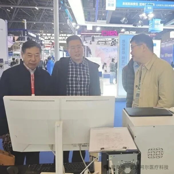
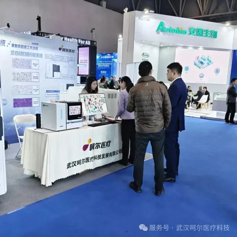
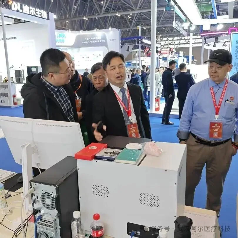
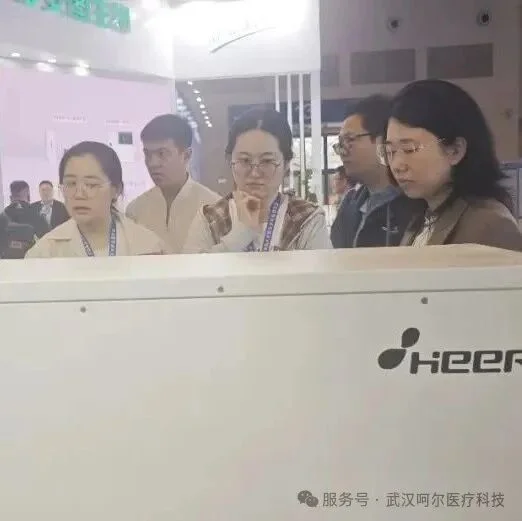
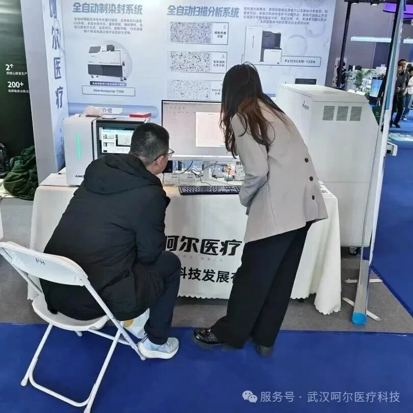
In late spring March, with the Wu Gorge stretching far and clouds casting floating light, from March 27 to 29, 2026, the "2026 Pathology Equipment (Chongqing) Seminar," organized by the Pathology Equipment Branch of the China Association of Medical Equipment and jointly hosted by the PLA Army Characteristic Medical Center (Daping Hospital) and the First Affiliated Hospital of Army Medical University (Southwest Hospital), was successfully held at the Chongqing International Expo Center. Heer Medical participated with its digital image scanning analysis system and liquid-based cytology pipeline, earning recognition from experts and customers alike.


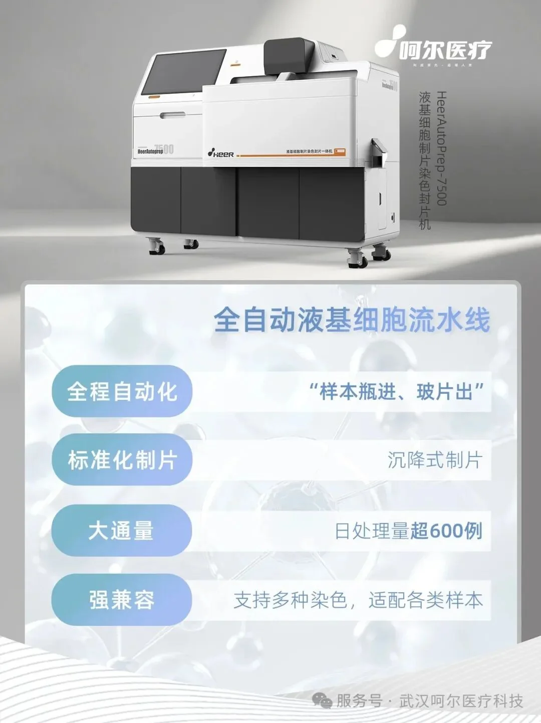
Liquid-Based Cytology Pipeline
Achieving Full-Process Automation, Empowering Digital Pathology Development
Heer Medical's self-developed liquid-based cytology pipeline and slide scanning image analysis system provide pathology departments with an integrated tumor screening solution. It effectively avoids the human variability and contamination risks inherent in traditional manual operations, producing thin-layer cell smears with uniform distribution and vivid colors. The "sample in, slide out" approach truly achieves full-process automation. With a daily processing capacity of over 600 samples, it significantly reduces operating time and labor costs. The standardized, high-quality slides produced through full-process automation also lay an ideal foundation for subsequent digital scanning and AI-assisted diagnosis.
Capable of completing independent preparation, staining, and coverslipping of 90 samples within one hour, with daily processing capacity exceeding 600 samples, boosting departmental work efficiency by 2 to 4 times.
Integrates information entry, automatic barcode scanning, automatic sample addition, and slide coding in one unit. Adopts a fully automated cell pipeline operation mode with a built-in barcode scanner, supporting unidirectional or bidirectional connection to departmental LIS/HIS systems.
Equipped with automatic sorting, one-time sedimentation chamber loading, and slide and coverslip loading functions, requiring no manual intervention. Can place 50 samples at once, truly achieving full-process automation of "sample in, slide out."
Supports both Pap staining and HE staining, suitable for preparation and staining of various samples including cervical exfoliated cells, serous cavity effusions, sputum, urine, and puncture fluids.
Uses natural sedimentation preparation, with the equipment simulating the real manual staining immersion process. After preparation, slides are automatically immersed in absolute ethanol and xylene for transparency, and wet coverslipping is automatically completed. Environmentally friendly transparency agents can also be used as alternatives to xylene. The equipment features a built-in exhaust system for safe and efficient operation. The resulting slide samples have evenly distributed cells with vivid colors, fully meeting the quality requirements for AI diagnosis.
Slide Scanning Image Analysis System
Both a Scanner and an Intelligent Analysis System
Heer Medical's self-developed slide scanning image analysis system integrates high-definition automated scanning with intelligent diagnosis, serving as both a scanner and an intelligent diagnostic system. The equipment is compact, fully functional, produces clear scan images, has a built-in scanning workstation, is easy to operate, and is ready to use upon installation.
Slide Scanning Image Analysis System
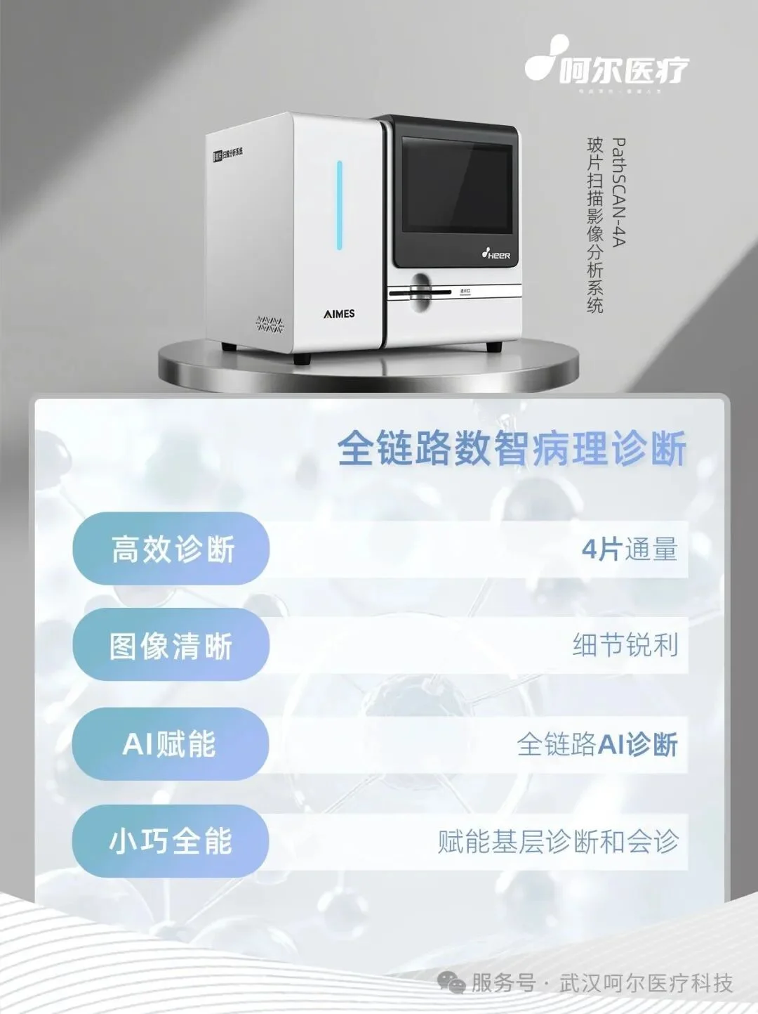
Full-Scenario AI Diagnostic Functions:
Used for scanning and observation of ordinary stained tissue sections, cell smears, and body fluid smears.
Used for DNA ploidy quantitative analysis of liquid-based cell samples (cervical, pleural/ascitic fluid, sputum, urine, and other exfoliated cells);
Used for AI morphological analysis of cervical cells;
Used for p16/Ki-67 dual-stain immunoanalysis;
Used for HPV E6/E7 protein detection;
Heer Medical is committed to empowering pathology departments in their digital and intelligent transformation, helping pathological diagnosis enter a new era of precision and efficiency.